|
|
|
|
|
|
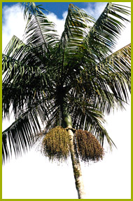
Stem
|
|
|
|
|
|
Straight, cylindrical, monopodial, single without building stolons at the base; smooth, grey, the stem is not considered bole. The palm reaches heights between 5 to 20 m and diameters between 8 and 30 cm (CARVALHO 1993). At the apex, place of leaf insertion, there is a smooth, green-olive to dark-green section of 1.0-1.5 m, thicker than the trunk, formed by a bunch of leaves. Within this section is the edible part of the palm.
|
|
|
|
|
|
|
|
|
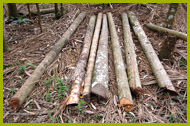
Stem of a circ. 40 year old E. edulis plant
|
|
|
|
Wood
|
|
|
|
Like the other Monocotyledons, the palm does not have a vascular cambium (meristem tissue layer between wood and bast that produces new cells), which causes the typical secondary growth. Thus, the growth in thickness is merely based on the expansion of existing cells. In the strict sense, if hereby by the cambium inwards secluded permanent tissue of Gymnospermae and Dikotyledonen is designated, palms do not have wood. But in its technological features and terms of lignification of the cell structures, the palm tissue is comparable with wood and will therefore be characterized as such (GROSSER 1977).
In contrast to the wood of conifers and hardwoods, the inside stem region of palm trees is always brighter than the outside. This difference is caused by groups of shaded cells, the so called vascular bundles, visible mainly in cross section. These bundles are immersed in a clear thin-walled tissue (parenchyma) and give the stem a great rigidity and stability. Due to the columnar growth of the stem, the palm wood is not homogeneous, but on the static point of view more solid on the outside and softer on the inside. Like a tube the soft inner core is surrounded by a hard outer sheath. As their main vascular system is not located in the outer area of the stem, the palm is excellently protected against fires.
|
|
|
Anatomical structure of the palm wood:
|
|
|
|
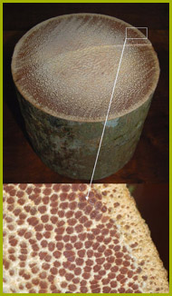
Macroscopically
(visible with the naked eye or at least magnifier)
|
|
|
 |
|
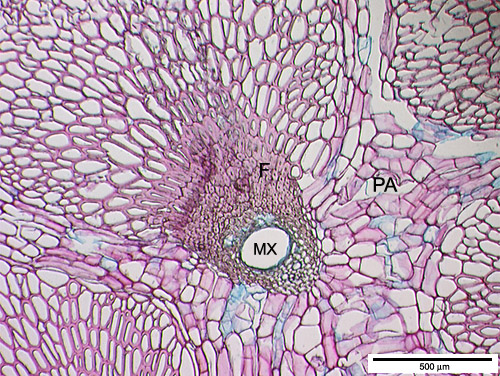
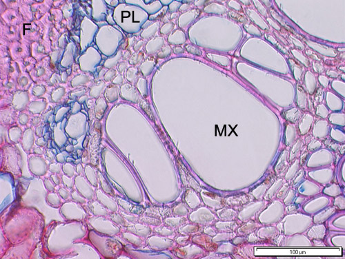
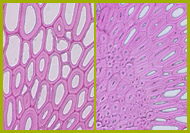
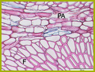
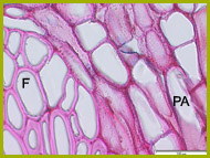
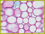
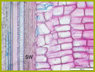
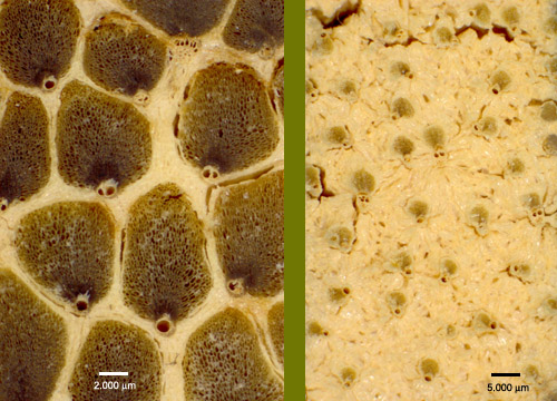
Macroscopic magnification of vascular bundles. Left: wide vascular bundle of the outer stem area with a high proportion of thick-walled, dark brown fibre cells as well as one or two Metaxylems. Right: small vascular bundles of the inner stem area, few in number and embedded in the bright parenchymatous ground tissue
|
|
|
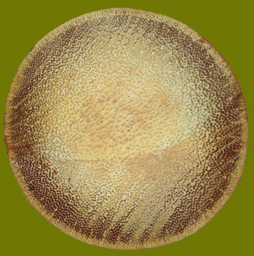
Visible in the cross-section of a palm stem are numerous scattered arranged black dots, the vascular bundles. Their number and dimension is decreasing strongly towards the stem centre. Beside this the brighter parenchymatous ground tissue and the small, a few millimetres thick bark is well-defined.
|
|
|
|
|